MASLD-associated gene variants
In the last 20 years, genome-wide association studies (GWAS) have identified common single-nucleotide polymorphisms in a variety of genes as main determinants of MASLD.
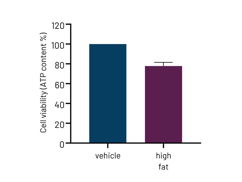
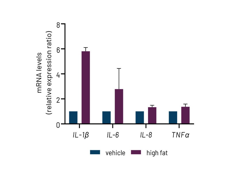
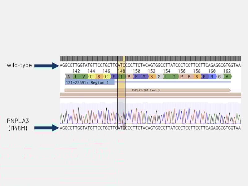
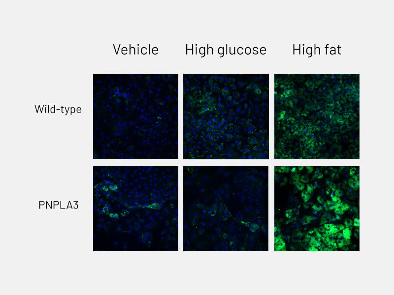
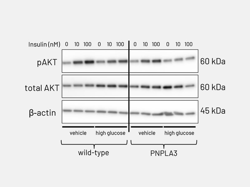
Of those, the most common disease-implicated genetic variant is I148M in the gene coding for Patatin-like phospholipase domain-containing protein 3 (PNPLA3). Using CRISPR/Cas9 technology, we have generated gene-edited Ulti-HEP models carrying the I148M mutation and demonstrated the detrimental effects of genetic variation on MASLD development.
This cell line is ideally suited for researchers who are specifically interested in modelling or screening compounds against hepatocytes with the PNPLA3 mutation. As with our wildtype Ulti-HEP cells, this mutated cell line is human-derived and source from a single donor, allowing you to scale your research as necessary whilst maintaining no batch-to-batch variation between cells.


.jpg)